
Tumors, both benign and malignant, can grow anywhere in the body. Our brain can also suffer from different types of tumors. One of these types is a meningioma.
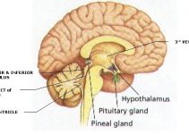
Our brain and spinal cord are covered by three different layers inside the skull and bony structures for protection and nourishment. These layers are called meninges.
Any tumor of meninges is called meningioma. These tumors can be benign or malignant. Most tumors are benign and involve the brain layers more commonly as compared to spinal cord layers. [1]
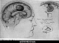
Table of Contents
What Causes A Meningioma?
There are multiple factors that can lead to the development of a meningioma. The most important risk factors are listed below.
- Exposure to radiation is one of the most important causes of developing meningioma. It affects children more adversely as compared with adults.
- Some nervous system diseases, such as neurofibromatosis, increase the risk of developing meningioma.
- Female sex is associated with a higher risk of having a meningioma. It is mainly because of female hormones.
- Obesity is a leading cause of many diseases. It is also found to be related to a higher meningioma risk. [1]
What are Different Grades of Meningioma?
Like any other type of tumor, meningioma also has various grades. Grading of a tumor is carried out to show the intensity of the disease. Meningioma is divided into three grades as below.
- Grade I is a slow-growing tumor that does not spread to other structures. It is benign.
- Grade II is a relatively quick growing tumor. Due to this, it is also referred to as atypical meningioma.
- Grade III is the most dangerous type. It grows rapidly and spreads to the other parts of the body as well. It is a malignant tumor.
Most meningiomas are grade I and can be treated. Grade II and III meningioma are difficult to treat but luckily, they are less prevalent. [2]

What are the Symptoms of Meningioma?
Meningioma can present with a wide variety of symptoms. These symptoms start gradually and become severe with time. The presentation usually depends on the area of involvement.
The symptoms are mainly caused due to the compression of the surrounding structures and an increase in the pressure inside the head.
The following are the most commonly experienced symptoms.
- Headache
- Nausea and vomiting
- Problem with vision like blurred vision
- Hearing loss
- Ringing sounds in ears
- A weakness of any part of the body
- Facial weakness
- Memory problems
- Concentration problems
- Confusion
- Loss of smell
- Seizures
The severity of symptoms usually depends on the size of the tumor. The smallest tumors sometimes do not produce any symptoms at all.
The frequency and the severity of symptoms increases as the size of the tumor increases. [3]
Another important fact about meningiomas is that sometimes they can run in families.
It is especially true for people with neurofibromatosis. It is a condition caused by genetic problems that run in family and it is associated with many types of tumors including meningioma.
So, people who have neurofibromatosis or meningioma in a family should be alert about the above-mentioned symptoms. [4]
Are Meningiomas Dangerous?
Dangers associated with meningioma depend on many factors, such as size, site, and type of tumor. Following complications can be faced in the case of a meningioma.
- Permanent disability
- Personality changes
- Memory and concentration problems
- Seizures
These problems can severely affect the quality of life. [3]
Survival Rates
The survival rates for malignant and non-malignant or benign meningioma are different. According to statistics, the ten-year survival rate for benign meningioma is 84%, while the ten-year survival rate for malignant meningioma is 62%. [5]
Surgical removal of a meningioma depends on many factors. These factors are considered before deciding on surgery for tumor removal. The following are the most important factors that are taken into account for this decision.
- Location of the tumor and symptoms it is producing
- If the tumor is benign or malignant
- How is the patient’s health and if he or she is suffering from any other health conditions
- Patient’s decision about treatment
Some benign and small tumor does not need any treatment at all. They are usually slow-growing tumors that do not produce any significant symptoms or complications.
However, your doctor monitors the tumor regularly and treats it when required. Studies show that even some small tumors can start enlarging at once leading to severe symptoms, so they should not be overlooked completely. [6]
The decision of removing a meningioma is also taken accordingly. The prognosis of a meningioma depends on its severity. Patients with a benign meningioma survive longer than people with aggressive or malignant meningioma.
What are Different Types of Meningioma?
Meningiomas are classified in different ways, such as on the basis of severity, location, and histology. The grading of meningiomas has already been discussed above.
The following are the names of meningioma that fall under histological grades I, II and III.
WHO grade I
- Meningothelial (syncytial) meningioma
- Transitional (mixed) meningioma
- Fibroblastic (fibrous) meningioma
- Psammomatous meningioma
- Angiomatous (vascular) meningioma
- Microcystic meningioma
- Secretory meningioma
- Lymphoplasmacyte-rich meningioma
- Metaplastic meningioma
WHO grade II
- Chordoid meningioma
- Clear-cell meningioma (intracranial)
- Atypical meningioma
WHO grade III
- Papillary meningioma
- Rhabdoid meningioma
- Anaplastic (malignant) meningioma
Grade I meningiomas are benign, grade II is moderately aggressive and grade III are aggressive or malignant meningiomas. [7]
Small tumors are usually less than 3cm in size. Tumors greater than 3 cm in size are considered big tumors. [2] Small-sized meningiomas are sometimes asymptomatic and the patient can have a nearly normal life with them.
The key is to follow-up regularly and to undergo regular imaging to ensure that there is no alarming increase in size or any other problem.
How is Meningioma Treated?
There are many treatment options for meningioma. They are listed below.
- Surgical resection is the most commonly adopted method.
- Radiotherapy is often carried out to decrease the size of a tumor or to treat residual tumors after surgery.
- Chemotherapy involves giving some medications that are most effective in aggressive tumors. This is the least commonly used treatment option.
However, surgery is the mainstay of the treatment of meningioma. Other options are considered if surgery is not possible due to various reasons, such as a deep location of tumor or patient’s bad general health.
These options can also be utilized along with surgery if complete surgical resection of the tumor is not possible due to its close proximity to important structures like blood vessels and nerves. [8]
There are different approaches that are utilized for meningioma resection.
The most common approaches are discussed below.
- A craniotomy involves opening the skull by making an incision. This approach is used to remove tumors that are present on the surface of the brain.
- The endoscopic endonasal approach involves entering the brain through the nose. A camera is introduced via the nose to visualize the tumor and then small instruments also enter the brain through the nose for the surgery.
- Neuroendoscopic approach is also a minimally invasive surgery in which cameras and instruments are introduced via small holes in the skull and surgery is performed.
- Spinal surgery is carried out for meningiomas involving the spinal cord.
The recovery period after a meningioma surgery varies from person to person and greatly depends on factors like the patient’s general health and how extensive the surgery was. It usually takes from a few days to a few weeks. [9]
All meningiomas are unique and have different behavior. Benign meningiomas are sometimes curable. There are high chances of recurrence even after a meningioma is completely treated.
So, although a benign meningioma can be cured, recurrence can occur.
Leaving a meningioma untreated is not an option in most cases. Some benign and slow-growing tumors are not treated aggressive but surveillance is vital to ensure patient’s safety.
If the patient is not followed up regularly, then the tumor may start growing quickly at some stage leading to permanent disability and other complications described above.
Some patients might not want to undergo surgery or the risk of surgery is high, so neurosurgeon decides to avoid surgery. In such cases, radiotherapy and chemotherapy are adopted for treatment.
However, the evidence says that surgical removal is the best treatment option, and radiotherapy and chemotherapy are not as effective as surgery. Chemotherapy is the least effective treatment option. [10]
Conclusion
Meningioma is a type of brain tumor that involves the outer layers of the brain and spinal cord. It has different grades ranging from slow-growing, benign masses to high-grade, aggressive malignant tumors.
It can produce various bothersome symptoms and can finally cause permanent disabilities and seizures.
The mainstay of treating a meningioma is surgical removal. However, radiotherapy and chemotherapy are also used as adjuvants or sometimes as primary treatment options when surgery is not possible due to various reasons.
They are not as useful as surgery. Even after surgery, meningiomas have a high rate of recurrence.
In some cases, a watchful wait is done before surgery is performed. It is done in benign tumors that slowly increase in size over the years. Regular follow-up is carried out.
Over time, surgery remains the major treatment modality for meningiomas. Surgery is carried out in most cases and it has a good outcome. Meningioma has a good survival rate if treated efficiently.
References
- https://www.ncbi.nlm.nih.gov/pmc/articles/PMC2945461/
- https://www.ncbi.nlm.nih.gov/pmc/articles/PMC5062955/
- https://www.ncbi.nlm.nih.gov/pmc/articles/PMC6123887/
- https://www.ncbi.nlm.nih.gov/pmc/articles/PMC6235681/
- https://www.aans.org/en/Patients/Neurosurgical-Conditions-and-Treatments/Meningiomas
- https://www.ncbi.nlm.nih.gov/pmc/articles/PMC5612578/
- https://www.ncbi.nlm.nih.gov/pmc/articles/PMC4078607/
- https://www.ncbi.nlm.nih.gov/pubmed/14732889
- https://www.ncbi.nlm.nih.gov/pmc/articles/PMC5879592/
- https://www.ncbi.nlm.nih.gov/pmc/articles/PMC4994143/